orals 5th Asia-Pacific NMR Symposium 2013
Oscillating Gradient Diffusion MRI in the ex-vivo Prostate: Regional Investigation of ADC Dependency on Gradient Oscillation Frequency (#64)
Introduction: Due to its sensitivity to subtle changes in cellular microstructure, DWI is a promising approach to improve diagnostic capabilities, particularly in pathologies as prostate cancer, that hardly show any changes in anatomical MRI. However, due to hardware restrictions, standard (pulsed-gradient) DWI methods are limited in resolving short diffusion times while maintaining sufficient sensitivity. Sampled free diffusion lengths are typicaly much larger than cellular dimensions. One way to overcome these restrictions, are oscillating gradient (OGSE) methods recently introduced to MRI2 . Here we investigate the dependency of OGSE ADCs on gradient oscillation frequency to distinguish cancerous and healthy tissues in the human prostate.
Methods: On a 9.4T Bruker BioSpec94/20 an OGSE-C1 sequence with waveforms as described in [3] was implemented. b-value/gradient relations were calculated from analytical relationships as derived in [2 ]. Sequence and signal were verified by hardware simulations and water phantom. An ex-vivo prostate was prepared as described in [1]. An OGSE-series using b-values 200, 400, 600, 800 and “b0” for oscillation frequencies 50Hz, 100Hz, 150Hz and 200Hz (∆eff = 5ms, 2.5ms, 1.66ms, 1.25ms). For each frequency ADC-maps were calculated by pixelwise fitting of all b-values with a mono-exponential model.
Results: Frequency dependent prostate ADC-maps are shown in Fig 1. ADC/oscillation-frequency dependency for different prostate regions is shown in Fig.2. The relative depiction (Fig.2b) shows strong differences in relative ADC increase with frequency for different tissue types.
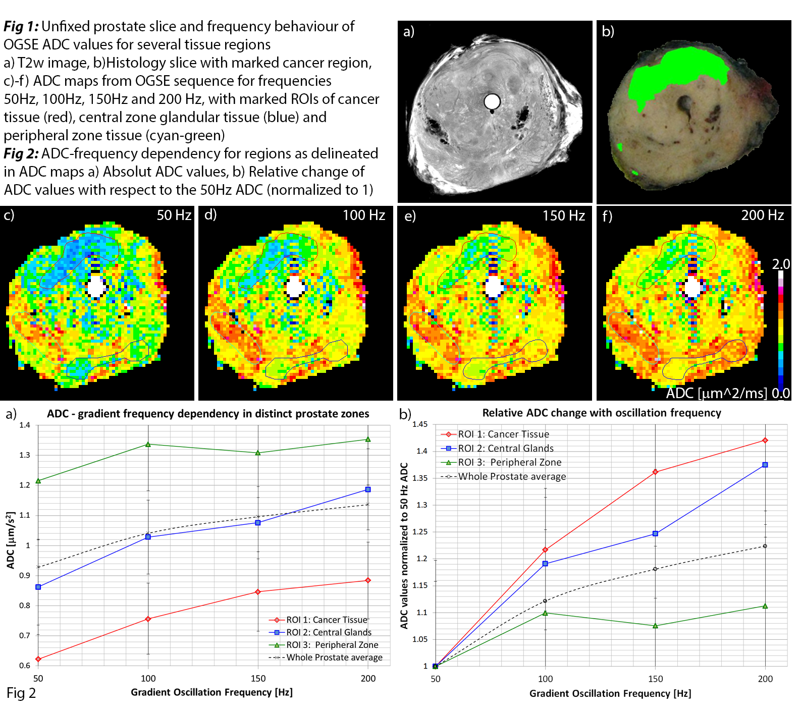
Conclusion: In the human prostate OGSE ADC-values show a strong dependency on gradient frequency (and hence on sampled diffusion time). Distinct tissue regions exhibit significantly different frequency behaviour, which might be exploited to distinguish prostate cancer from healthy tissues. Additional studies are needed to elucidate the details and cellular origins of these findings. Denser sampling of higher frequencies can enable theoretical OGSE diffusion modeling and determination of average cell sizes.
- Roger Bourne, Andre Bongers, Ned Charles, Carl Power, Paul Sved, and Geoffrey Watson, 'Effect of Formalin Fixation on Biexponential Modeling of Diffusion Decay in Prostate Tissue', Magnetic Resonance in Medicine (2012).
- Mark D Does, Edward C Parsons, and John C Gore, 'Oscillating Gradient Measurements of Water Diffusion in Normal and Globally Ischemic Rat Brain', Magnetic resonance in medicine, 49 (2003), 206-15.
- Edward C Parsons, Mark D Does, and John C Gore, 'Temporal Diffusion Spectroscopy: Theory and Implementation in Restricted Systems Using Oscillating Gradients', Magnetic resonance in medicine, 55 (2006), 75-84.