orals 5th Asia-Pacific NMR Symposium 2013
Longitudinal study of the diffusion tensor imaging of the corpus callosum in a rodent model of hydrocephalus. (#39)
Background: Hydrocephalus (HCP) is a neurological disease characterized by an enlargement of the cerebral ventricles resulting in compression of surrounding tissues(1, 2). The disease diagnosis does not necessarily match with ventricular enlargement, but can be related to the integrity of the brain tissue (3). In this context, additionally to quantify the ventriculomegaly, brain damage need to be evaluated by assessing changes in its micro-structure. As Diffusion Tensor Imaging (DTI) is capable of yielding information on axonal organization (4, 5), this technique can be used to investigate how this property may change with compression. For this reason, DTI was used in parallel with anatomical imaging to study early stages of HCP development in a longitudinal study.
Methods: HCP was induced in SD rats (female; 4 weeks old, n=5) by injecting kaolin into the cisterna magna. One saline-injected rat acted as a control. Magnetic Resonance (MR) imaging (9.4T, Bruker) was performed on the rats one day before the injection, and 3, 7 and 16 days post-injection (Fig.1). T2-weighted and DTI imaging (32 gradient directions, b-factor 670 s/mm2) were performed. Ventricle ratios were measured from anatomical images and the relationship with fractional anisotropy (FA) and mean diffusivity (MD) of the corpus callosum, as measured on the DTI images using DSI Studio, was evaluated using Spearman’s correlation.
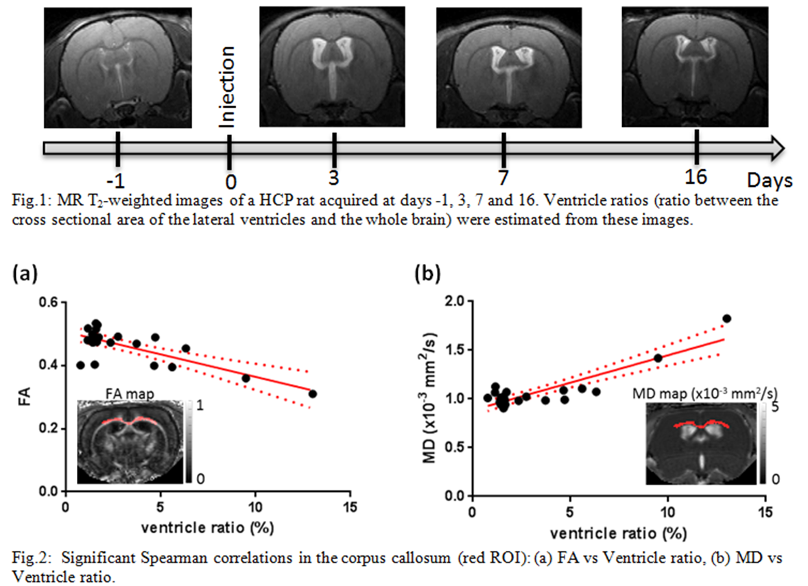
Results: As the ventricle ratio increased, FA decreased and MD increased (Fig.2). Both relationships were significant (r=-0.4555, 0.4160 / P=0.03, 0.048 respectively).
Conclusions: Changes in tissue diffusion properties in hydrocephalus are a potential method to evaluate the severity of the damage to brain tissue in HCP. This study presents important preliminary data on the development of new potential imaging biomarkers for this disease.
The authors would like to thank Sarah Hemley and Elmira Najafi for training in the surgical technique.
- Assaf, Y., Ben-Sira, L., Constantini, S., Chang, L.C. & Beni-Adani, L. (2006) Diffusion tensor imaging in hydrocephalus: initial experience. AJNR Am J Neuroradiol, 27, 1717-1724.
- Del Bigio, M.R. (2010) Neuropathology and structural changes in hydrocephalus. Dev Disabil Res Rev, 16, 16-22.
- Weller, R.O. & Shulman, K. (1972) Infantile hydrocephalus: clinical, histological, and ultrastructural study of brain damage. J Neurosurg, 36, 255-265.
- Pierpaoli, C., Jezzard, P., Basser, P.J., Barnett, A. & Di Chiro, G. (1996) Diffusion tensor MR imaging of the human brain. Radiology, 201, 637-648.
- Pierpaoli, C. & Basser, P.J. (1996) Toward a quantitative assessment of diffusion anisotropy. Magn Reson Med, 36, 893-906.