posters 5th Asia-Pacific NMR Symposium 2013
Effects of deuterium oxide on the anistropy of proton spin relaxation in articular cartilage observed using MR micro-imaging (#209)
Background: Understanding interactions between water molecules and macromolecular components of cartilage allows quantitative assessment of its structure and composition. It is known that dipolar couplings in water molecules are weaker when protons are exchanged with deuterium nuclei1 . In this study, anisotropy of proton spin relaxation in partially deuterated articular cartilage was observed using MR micro-imaging experiments.
Methods: Four articular cartilage samples were excised from visually normal bovine patellae. T1- and T2-weighted imaging was performed at two sample orientations θAS= 0° and 55°, where θAS is the angle between the static magnetic field B0 and the normal to the articular surface of the sample. Each sample was soaked for at least 12 hours in four solutions with molar concentration ratios of %D2O:%H2O= 0:100, 25:75, 50:50, 75:25. The imaging protocol was repeated after each soaking session. Non-linear curve fitting yielded R1 and R2 relaxation rates for each voxel. Regions of interest encompassing cartilage were manually selected.
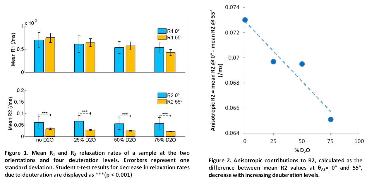
Results: Figure 1 displays R1 and R2 rates in one sample at each D2O concentration at the two orientations. Longitudinal relaxation is isotropic, while transverse relaxation is anisotropic; that is, there is significant difference between R2 relaxation rates at the two orientations. This difference can be attributed to the anisotropic contributions to relaxation, caused by residual dipolar couplings in water molecules bound to aligned collagen fibers2,3, which are minimized at the magic angle. It was also found that these anisotropic contributions decreased with increasing D2O concentrations, as shown in Figure 2. However, both the composition and the collagen alignment vary across the cartilage and further investigations, preferably based on voxel-by-voxel comparisons, are necessary.

Conclusion: Deuterium oxide was used to modify the anisotropic contributions to relaxation. This is useful in investigations of proton relaxation mechanisms in cartilage.
- Shapiro, E. M., Borthakur, A., et al. Osteoarthritis and cartilage 9, 533–8 (2001).
- Xia, Y. Magnetic Resonance in Medicine 39, 941–949 (1998).
- Momot, K. I., Pope, J. M. & Wellard, R. M. NMR in biomedicine 23, 313–24 (2010).